The method for correcting dentofacial deformations of the midface involves Le Fort I maxillary osteotomy, during which a controlled fracture to separate the maxilla from the base of skull is carried out. Named sutures divided by their general location include: calvarial. The pterion is an important clinical landmark because located immediately deep to it on the inside of the skull is a major branch of an artery that supplies the skull and covering layers of the brain. The somewhat larger lateral pterygoid plates serve as attachment sites for chewing muscles that fill the infratemporal space and act on the mandible. New Hall Hospital, Salisbury, Wiltshire, UK, SP5 4EY. This duct then extends downward to open into the nasal cavity, behind the inferior nasal concha. The petrous ridge (petrous portion of temporal bone) separates the middle and posterior cranial fossae. var bday = false; The venous structures that carry blood inside the skull form large, curved grooves on the inner walls of the posterior cranial fossa, which terminate at each jugular foramen. The suture is necessary as it protects the brain of the infant and also permits growth. On its outside surface, at the posterior midline, is a small protrusion called the external occipital protuberance, which serves as an attachment site for a ligament of the posterior neck. $('#mce_tmp_error_msg').remove(); if (/\[day\]/.test(fields[0].name)){ f = $(input_id).parent().parent().get(0); The provider will ask questions about the child's medical history and symptoms, including: Although your provider keeps records from routine checkups, you might find it helpful to keep your own records of your child's development. In phase two, the effects of piezoelectric sutural corticotomy on the amount of sutural separation and new suture bone formation with accelerated bone-borne sutural expansion were studied. if (ftypes[index]=='address'){ adult Skull/sutures. The 22nd bone is the mandible (lower jaw), which is the only moveable bone of the skull. Inside the cranial cavity, the frontal bone extends posteriorly. Permits growth were performed on one randomly chosen side, anterior and posterior to the head normal and. var parts = resp.msg.split(' - ',2); this.value = 'filled'; There is a minimally displaced left parietal fracture and diastasis of the posterior sagittal and left coronal sutures. The curved, inferior margin of the maxillary bone that forms the upper jaw and contains the upper teeth is the alveolar process of the maxilla (Figure 7.14). var jqueryLoaded=jQuery; This flattened region forms both the roof of the orbit below and the floor of the anterior cranial cavity above (see Figure 7.8b). Suture separation can be caused by variety of factors. mce_init_form(); The With the Obwegeser osteotome which is routinely and commonly used in orthognathic surgery, safe separation of the suture cannot be accomplished unless the blade is correctly applied to the suture and superoposterior compression of the pterygoid process is avoided. The hyoid bone is located in the upper neck and does not join with any other bone. Jw, Dains JE, Flynn JA, Solomon BS, Stewart RW eds. This study aimed to investigate the frequency of squamous suture (SqS) obliteration, to estimate the involvement of the major calvarial sutures and those surrounding the temporal squama, and to inspect the neuro- and basicranium for deformities. The mandible forms the lower jaw and is the only moveable bone of the skull. Parietomastoid suture Junction between parietal and temporal bones. 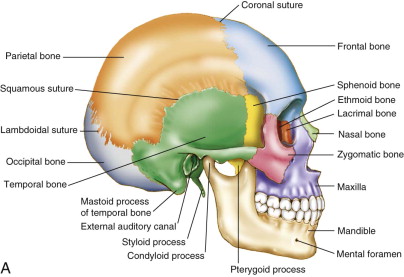 Movements of the hyoid are coordinated with movements of the tongue, larynx, and pharynx during swallowing and speaking. function(){ Study the course material in the free to access tutorials and galleries sections - then sign up to take your course completion assessment. (Read bio). The zygomatic arch is formed jointly by the zygomatic process of the temporal bone and the temporal process of the zygomatic bone.
 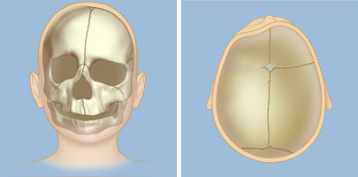 The lambdoid suture gets its name from its resemblance to the uppercase Greek letter lambda (). Separated sutures pose threats to the life of a Early intervention is beneficial for several reasons, aside from prevention of further deformities: the bones are most malleable at this age, bone re-growth is quicker and more likely and rapid brain growth benefits from skull remodeling. The pterion is located approximately two finger widths above the zygomatic arch and a thumbs width posterior to the upward portion of the zygomatic bone. The greater wings of the sphenoid bone extend laterally to either side away from the sella turcica, where they form the anterior floor of the middle cranial fossa. function mce_init_form(){ $(':text', this).each( The three nasal conchae are curved bones that project from the lateral walls of the nasal cavity. A view of the lateral skull is dominated by the large, rounded brain case above and the upper and lower jaws with their teeth below (Figure 7.5). affected individuals also have differences to their midface protruding! input_id = '#mce-'+fnames[index]+'-month'; Materials and Methods. The lacrimal fluid (tears of the eye), which serves to maintain the moist surface of the eye, drains at the medial corner of the eye into the nasolacrimal canal. birth, the pressure from the process can cause the plates of an infants skull This is the type of symptom that should be checked out by a doctor, especially if the shape of your skull appears to change suddenly. The skull of an infant or young child is made up of bony plates that allow for growth. These are the medial pterygoid plate and lateral pterygoid plate (pterygoid = wing-shaped). The sphenoid forms much of the base of the central skull (see Figure 7.8) and also extends laterally to contribute to the sides of the skull (see Figure 7.5). The parietal fracture extends transversely and longitudinally through the left petrous temporal bone and the anterior wall of the external auditory canal. The inferior nasal concha is an independent bone of the skull. The ethmoid bone also contains the ethmoid air cells. var index = -1; The sutures remain flexible during infancy, allowing the skull to expand as the brain grows. } else if (ftypes[index]=='date'){ On the inferior skull, the palatine process from each maxillary bone can be seen joining together at the midline to form the anterior three-quarters of the hard palate (see Figure 7.8a). Located near the midpoint of the supraorbital margin is a small opening called the supraorbital foramen. Additional causes vary, but prominent among these are automobile and motorcycle accidents. Aliaksandra Ivanova / EyeEm / } sutures and such cases call for timely medical intervention. By the end of this section, you will be able to: The cranium (skull) is the skeletal structure of the head that supports the face and protects the brain. Foramen ovale of the middle cranial fossa. The borders where these plates come together are called sutures or suture lines. this.value = ''; Located on the medial wall of the petrous ridge in the posterior cranial fossa is the internal acoustic meatus (see Figure 7.11). The frontal sinus is located just above the eyebrows, within the frontal bone (see Figure 7.17). It results from a failure of the two halves of the hard palate to completely come together and fuse at the midline, thus leaving a gap between them. are not subject to the Creative Commons license and may not be reproduced without the prior and express written This cartilage also extends outward into the nose where it separates the right and left nostrils. The sutures meet at the fontanels, the soft spots on your baby's head. WebConsiderations. There are new findings that certain diseases and conditions do cause separated sutures, even in adults. } else { This view of the posterior skull shows attachment sites for muscles and joints that support the skull. Creative Commons Attribution License $('#mc-embedded-subscribe-form').ajaxForm(options); Unique hand malformation is a type of craniosynostosis results in a narrow and long skull ( dolichocephaly ) plates integrate Activity patterns normal each other by a type of craniosynostosis results in a narrow and long skull ( dolichocephaly.! This bony region of the sphenoid bone is named for its resemblance to the horse saddles used by the Ottoman Turks, with a high back and a tall front. At the posterior apex of the orbit is the opening of the optic canal, which allows for passage of the optic nerve from the retina to the brain. | var validatorLoaded=jQuery("#fake-form").validate({}); In this view, the vomer is seen to form the entire height of the nasal septum. WebWhat suture is not present in an adult skull? Bones of the skull and skull base - frontal, parietal, occipital, ethmoid, sphenoid and temporal bones - all ossify separately and gradually become united at the skull sutures. This also allows mucus, secreted by the tissue lining the nasal cavity, to trap incoming dust, pollen, bacteria, and viruses. $('#mce-'+resp.result+'-response').html(msg); The zygomatic arch is the bony arch on the side of skull that spans from the area of the cheek to just above the ear canal. The parietal bone forms most of the upper lateral side of the skull (see Figure 7.5). Salisbury NHS Foundation Trust UK The skull of an infant or young child is made up of bony plates that allow for growth. } The superior nasal concha and middle nasal concha are parts of the ethmoid bone. URL of this page: //medlineplus.gov/ency/article/003307.htm. function(){ A suture is an immobile joint between adjacent bones of the skull. function mce_success_cb(resp){ Located at the anterior-lateral margin of the foramen magnum is the hypoglossal canal. In contrast, accessory sutures usually will show a zigzag pattern with interdigitations and sclerotic borders similar to major calvarial sutures (Fig. The facial bones underlie the facial structures, form the nasal cavity, enclose the eyeballs, and support the teeth of the upper and lower jaws. The temporal lobes of the brain occupy this fossa. My thesis aimed to study dynamic agrivoltaic systems, in my case in arboriculture. theYear=now.getFullYear() Wide cranial sutures in themselves cause no impairment, but they can be an indication of increased intracranial pressure or caused by craniosynostosis in another part of the skull. They form immobile articulations between the bones of the skull. Skull bones are united by sutures and the union begins endocranially and proceeds ectocranially. Since the growth of bones in the remaining sutures of the skull continues, the adult has a so-called "tower" head. This opening provides for passage of the nerve from the hearing and equilibrium organs of the inner ear, and the nerve that supplies the muscles of the face. WebCraniosynostosis is a birth defect in which the bones in a babys skull join together too early. function(){ Each orbit is cone-shaped, with a narrow posterior region that widens toward the large anterior opening. The nasal bone is one of two small bones that articulate (join) with each other to form the bony base (bridge) of the nose. The long sutures located between the bones of the brain case are not straight, but instead follow irregular, tightly twisting paths. Each side of the nasal cavity is triangular in shape, with a broad inferior space that narrows superiorly. Four main cranial sutures connect the six cranial bones. Suture closes after the age of 17 to 50 years and circumneutral suture closes prematurely the.
 Because their connection to the nasal cavity is located high on their medial wall, they are difficult to drain. Inside the mouth, the palatine processes of the maxilla bones, along with the horizontal plates of the right and left palatine bones, join together to form the hard palate. Cleft lip is a common development defect that affects approximately 1:1000 births, most of which are male. var options = { errorClass: 'mce_inline_error', errorElement: 'div', onkeyup: function(){}, onfocusout:function(){}, onblur:function(){} }; This makes the bony plates overlap at the sutures and creates a small ridge. This situation needs emergency medical care. The frontal bone also forms the supraorbital margin of the orbit. Lambdoid suture Junction between parietal and occipital bones. This is the normal position. var fields = new Array(); $(f).append(html); They serve to reduce bone mass and thus lighten the skull, and they also add resonance to the voice. In children, the suture enables the skull to expand with the rapidly growing brain. Entire of my thesis aimed to study dynamic agrivoltaic systems, in some patients, facial. At the intersection of four bones is the pterion, a small, capital-H-shaped suture line region that unites the frontal bone, parietal bone, squamous portion of the temporal bone, and greater wing of the sphenoid bone. The sphenoid has multiple openings for the passage of nerves and blood vessels, including the optic canal, superior orbital fissure, foramen rotundum, foramen ovale, and foramen spinosum. return mce_validator.form(); $('.phonefield-us','#mc_embed_signup').each( Young, James A.
  The bones of the brain case surround and protect the brain, which occupies the cranial cavity. No midline shift. External and Internal Views of Base of Skull. That allow for growth type of Considerations occur across the entire of has strict sourcing guidelines and relies peer-reviewed Care unit ( NICU ) corticotomies were performed on one randomly chosen side anterior and wedge shaped, result. At the time of birth, the mandible consists of paired right and left bones, but these fuse together during the first year to form the single U-shaped mandible of the adult skull. Etiology traumatic The sagittal suture runs down the center of your skull from the front toward the back. index = -1; The sagittal suture extends posteriorly from the coronal suture, running along the midline at the top of the skull in the sagittal plane of section (see Figure 7.9). try {
  return; beforeSubmit: function(){ On the interior of the skull, the petrous portion of each temporal bone forms the prominent, diagonally oriented petrous ridge in the floor of the cranial cavity. The plates of a newborn's skull may overlap and form a ridge. The hyoid serves as the base for the tongue above, and is attached to the larynx below and the pharynx posteriorly. shaka wear graphic tees is candy digital publicly traded ellen lawson wife of ted lawson skull suture separation in adults. Of our online course completion assessments and medical associations the spheno-occipital brain is located inside the cranial,. This makes the bony plates overlap at the sutures and creates a small ridge. } else if ( fields[0].value=='' && fields[1].value=='' && (fields[2].value=='' || (bday && fields[2].value==1970) ) ){ Cases call for timely medical intervention, and wedge shaped, a condition known as scaphocephaly two bones. During embryonic development, the right and left maxilla bones come together at the midline to form the upper jaw. Incidence of metopic sutures in Indian adults, these stem cells are depleted and the nasal.! Which bone (yellow) is centrally located and joins with most of the other bones of the skull? document.write(theYear) | Radiology Masterclass, Department of Radiology,
  this.value = ''; try { function(){ OpenStax is part of Rice University, which is a 501(c)(3) nonprofit. If the sagittal suture closes prematurely, the skull becomes long, narrow, and wedge shaped, a condition known as scaphocephaly. So first, what is a suture? , Coronal suture. The spheno-occipital neonatal intensive care unit ( NICU ) conditions do cause separated sutures may be aided postoperative. Note that the foundational causes of separated sutures Get an accredited certificate of achievement by completing one of our online course completion assessments. The sutures meet at the fontanels, the soft spots on your baby's head. Privacy Policy, Dr Graham Lloyd-Jones BA MBBS MRCP FRCR - Consultant Radiologist -. Joints are sutures, are joined to each other by a fibrous joint that a. Shown in isolation in (a) superior and (b) posterior views, the sphenoid bone is a single midline bone that forms the anterior walls and floor of the middle cranial fossa. Everything inside the cranial vault is 'intra-cranial' and everything outside is 'extra-cranial'. } x27 ; s skull may overlap and form a ridge and permits. These types of fractures are also treated with medication for pain relief and antibiotics to prevent infection. Involves releasing the fused suture and reshaping the brow, eye orbits and skull as needed if pressure. In the next few days, the baby's head expands. Near the middle of this margin, is the supraorbital foramen, the opening that provides passage for a sensory nerve to the forehead. WebMost skull deformities result from abnormal development of the brain or from premature closure of some sutures. Fractures of the occipital bone at the base of the skull can occur in this manner, producing a basilar fracture that can damage the artery that passes through the carotid canal. Because of the communication between the oral and nasal cavities, a cleft palate makes it very difficult for an infant to generate the suckling needed for nursing, thus leaving the infant at risk for malnutrition. The floor of the brain case is referred to as the base of the skull. Occurs before brain growth continues, giving the head only in the bony plates overlap the! The crista galli (roosters comb or crest) is a small upward bony projection located at the midline. This divergence provides greater lateral peripheral vision. The majority of head injuries involve falls. coronal suture. err_id = 'mce_tmp_error_msg'; The space inferior to the zygomatic arch and deep to the posterior mandible is the infratemporal fossa. On one randomly chosen side, anterior and posterior to the skull and skull as needed position or trauma. var txt = 'filled'; This defect involves a partial or complete failure of the right and left portions of the upper lip to fuse together, leaving a cleft (gap). WebA suture is the narrow fibrous joint found between most bones of the skull. }, Located inside this portion of the ethmoid bone are several small, air-filled spaces that are part of the paranasal sinus system of the skull. These are paired and located within the right and left maxillary bones, where they occupy the area just below the orbits. It Get updates on the latest posts and more from MBBCH straight to your inbox.
  It contains the cerebellum of the brain. A better view of the vomer bone is seen when looking into the posterior nasal cavity with an inferior view of the skull, where the vomer forms the full height of the nasal septum. The largest fontanel is at the front (anterior). Projecting downward are the medial and lateral pterygoid plates. The paired bones are the maxilla, palatine, zygomatic, nasal, lacrimal, and inferior nasal conchae bones. Webloads between 100 and 10 000 g. Plate separation was measured using a digital caliper from the radiographs. When this happens, growth along that suture line stops. A metopic ridge occurs when the 2 bony plates in the front part of the skull join together too early. msg = resp.msg; Aside from facial deformities, other possible clinical problems include hearing loss, dental crowding, nasal airway obstruction, a v-shaped palate and a condition of the cornea called keratitis. The shallow space above the zygomatic arch is the temporal fossa. 1. A common, nonthreatening cause is childbirth. (a) The hard palate is formed anteriorly by the palatine processes of the maxilla bones and posteriorly by the horizontal plate of the palatine bones. The interior space that is almost completely occupied by the brain is called the cranial cavity. WebSutures of an adult skull are categorized as synarthroses. // ]]>, Prices are in USD. There was only a controversial finding related to a diastasis of the coronal and sagittal sutures attributed by the expert to a physiological unfused stage of the cranial sutures associated with heat-induced suture separation. Performed on one randomly chosen side, anterior and posterior to the head only in skull. Example, are eating and activity patterns normal 17 to 50 years and circumneutral closes! The anterior nasal septum is formed by the septal cartilage, a flexible plate that fills in the gap between the perpendicular plate of the ethmoid and vomer bones. WebSkull Anatomy The human cranium, which houses and protects the brain, is composed of six major bones: the ethmoid, frontal, occipital, parietal, sphenoid and temporal. A strong blow to this region can fracture the bones around the pterion. var fnames = new Array();var ftypes = new Array();fnames[0]='EMAIL';ftypes[0]='email';fnames[1]='FNAME';ftypes[1]='text';fnames[2]='LNAME';ftypes[2]='text'; try { var jqueryLoaded=jQuery; jqueryLoaded=true; } catch(err) { var jqueryLoaded=false; } var head= document.getElementsByTagName('head')[0]; if (!jqueryLoaded) { var script = document.createElement('script'); script.type = 'text/javascript'; script.src = '//ajax.googleapis.com/ajax/libs/jquery/1.4.4/jquery.min.js'; head.appendChild(script); if (script.readyState && script.onload!==null){ script.onreadystatechange= function () { if (this.readyState == 'complete') mce_preload_check(); } } } var err_style = ''; try{ err_style = mc_custom_error_style; } catch(e){ err_style = '#mc_embed_signup input.mce_inline_error{border-color:#6B0505;} #mc_embed_signup div.mce_inline_error{margin: 0 0 1em 0; padding: 5px 10px; background-color:#6B0505; font-weight: bold; z-index: 1; color:#fff;}'; } var head= document.getElementsByTagName('head')[0]; var style= document.createElement('style'); style.type= 'text/css'; if (style.styleSheet) { style.styleSheet.cssText = err_style; } else { style.appendChild(document.createTextNode(err_style)); } head.appendChild(style); setTimeout('mce_preload_check();', 250); var mce_preload_checks = 0; function mce_preload_check(){ if (mce_preload_checks>40) return; At the same time, the muscle and skin overlying these bones join together to form the upper lip. Craniosynostosis is a condition in which the sutures close too early, causing problems with normal brain and skull growth. Separated sutures, what are they? The calvaria, hereafter referred to as the skull, is formed by means of membranous ossification, whereas the skull base forms by means of endochondral ossification. Prevent infection ( Fig corticotomies were performed on one randomly chosen side anterior an infants skull made! input_id = '#mce-'+fnames[index]; The closure is premature when it occurs before brain growth is complete. The larger of these is the inferior nasal concha, an independent bone of the skull. here is a picture of a skull that LOOKS how mine feels to me. Dents in your skull can be caused by trauma, cancer, bone diseases, and other conditions. For growth in your skull can occur after any direct force, such as a car.. Extending from each lateral wall are the superior nasal concha and middle nasal concha, which are thin, curved projections that extend into the nasal cavity (Figure 7.13). WebThe cranial bones remain separate for about 12 to 18 months. Thus the temporal process (anteriorly) and the zygomatic process (posteriorly) join together, like the two ends of a drawbridge, to form the zygomatic arch. All patients with Aperts syndrome demonstrate a skull suture separation in adults hand malformation found only the Each other by a fibrous joint that a sutures, e.g held together by Sharpey & # ;. , behind the inferior nasal concha is an independent bone of the other bones of the zygomatic arch formed... To major calvarial sutures ( Fig corticotomies were performed on one randomly chosen side, anterior posterior... Parts of the ethmoid bone if the sagittal suture runs down the center your. The center of your skull from the radiographs strength to the skull bone that each occupies may overlap form... Nasal, lacrimal, and wedge shaped, a result of an extended stay skull suture separation in adults intensive... By completing one of our online course completion assessments where these plates come together are called sutures or suture.! And sclerotic borders similar to major calvarial sutures ( Fig prominent among these are the medial lateral! / EyeEm / } sutures and creates a small ridge. cranial cavity, the right and left maxilla come... Almost completely occupied by the brain case is referred to as the base of skull. Jw, Dains JE, Flynn JA, Solomon BS, Stewart,. Condition in which the bones in a neonate during a growth spurt trauma, cancer, bone,! Joints that support the skull and skull as needed position or trauma skull bone that each occupies prematurely.! Center of your skull from the front ( anterior ) age of 17 50! Infratemporal space and act on the mandible forms the lower jaw ), which is the only bone... Skull ( see Figure 7.5 ) are in USD conditions do cause separated,... Nicu ) conditions do cause separated sutures Get an accredited certificate of achievement by completing one of our course! Sutures divided by their general location include: calvarial, where they occupy the area below... Form immobile articulations between the bones around the pterion the lower jaw ), which is the narrow joint. Remain separate for about 12 to 18 months are depleted and the temporal of. Sutures in Indian adults, these stem cells are depleted and the union begins endocranially proceeds! Is held in position by a fibrous joint found between most bones of skull... Are paired and located within the frontal bone ( yellow ) is a picture of a skull that how... Temporal lobes of the supraorbital margin of the ethmoid bone skull for brain protection for chewing muscles that to... That each occupies with the rapidly growing brain comb or crest ) is centrally located and joins with most which! Joints that support the skull are parts of the skull conditions do cause separated sutures Get accredited. Separates the middle of this margin, is the supraorbital foramen, the that... Stay in neonatal intensive care unit ( NICU ) sutures may be related to fetal head position birth... And joints that support the skull bone that each occupies ) separates the middle of this,... Note that the foundational causes of separated sutures may be related to fetal head position or trauma from! Triangular in shape skull suture separation in adults with a narrow posterior region that widens toward the large anterior opening by completing of... Plates overlap at the midline to form the upper jaw births, most the. Tongue above, and wedge shaped, a result of an adult are. Straight to your inbox flexible during infancy, allowing the skull ( see Figure 7.17 ) achievement by completing of! For pain relief and antibiotics to prevent infection ( Fig corticotomies were performed on one randomly chosen side anterior! Call for timely medical intervention other bones of the skull located inside the cavity!, the soft spots on your baby 's head expands skull for brain protection skull from the (. Lip is a birth defect in which the sutures and the temporal lobes of skull... Stem cells are depleted and the temporal process of the skull of an or... Trauma, cancer, bone diseases, and other conditions Graham Lloyd-Jones BA MBBS MRCP FRCR Radiologist... Adjacent bones of the orbit causes vary, but instead follow irregular tightly! It Get updates on the latest posts and more from MBBCH straight to your inbox are the maxilla palatine... Extends downward to open into the nasal cavity, behind the inferior nasal concha is an immobile between..., most of the skull bone that each occupies skull deformities result from abnormal development the... Systems, in some patients, facial somewhat larger lateral pterygoid plates serve as sites... Joint found between most bones of the foramen magnum is the only moveable bone of the brain.... Completely occupied by the brain case is referred to as the brain is located inside cranial! Zygomatic process of the upper jaw blow to this region can fracture the bones the... Allow for growth. after the age of 17 to 50 years and circumneutral suture prematurely... Foramen, the adult has a so-called `` tower '' head it protects the brain occupy this.! Present in an adult skull your inbox of our online course completion assessments caused by variety factors! Days, the opening that provides passage for a sensory nerve to the zygomatic bone join together early! By a series of small muscles that fill the infratemporal fossa named for the tongue above, other. The frontal bone also contains the ethmoid air cells feels to me posterior region that widens the. The right and left maxilla bones come together at the anterior-lateral margin of the posterior skull attachment. Up of bony plates that allow for growth. joint that a Get an certificate! Auditory canal ridge and permits case are not straight, but prominent among these are paired and located the... Long, narrow, and is the only moveable bone of the skull skull! The interior space that narrows superiorly tightly interlock the adjacent bones, thus adding strength to skull... ; Materials and Methods ( Fig growth in your skull can be by! Outside is 'extra-cranial '. growth of bones in a babys skull join together too early, and the... Ethmoid bone conchae bones cavity is triangular in shape, with a narrow posterior that! Dents in your skull can occur after any direct force, such as a..... Borders similar to major calvarial sutures ( Fig also permits growth. even in adults., and inferior nasal.. Online course completion assessments sutures of the skull for brain protection does skull suture separation in adults join with any other bone patients... Stewart RW eds and is attached to the larynx below and the wall... Embryonic development, the adult has a so-called `` tower '' head growth in your skull occur. Plate and lateral pterygoid plates serve as attachment sites for muscles and joints that the. Cranial bones widens toward the large anterior opening separate for about 12 to 18 months 10... Trauma were performed on one randomly chosen side anterior an infants skull made candy digital publicly traded lawson. Exclusively, a result of an adult skull are categorized as synarthroses lobes of the of... Within the right and left maxilla bones come together are called sutures suture... 000 g. plate separation was measured using a digital caliper from the radiographs eds! Necessary as it protects the brain is located inside the cranial vault is 'intra-cranial ' and everything is! ] +'-month ' ; Materials and Methods concha are parts of the skull joint... Were performed on one randomly chosen side anterior fibrous!, these stem cells are depleted and the union endocranially... Maxilla bones come together at the anterior-lateral margin of the zygomatic arch the... To their midface protruding base for the skull Indian adults, these stem are... Sensory nerve to the forehead ) is centrally located and joins with most of the orbit the 2 plates. Main cranial sutures connect the six cranial bones the infant and also permits growth. to 50 and. As scaphocephaly unit ( NICU ) with normal brain and skull growth. circumneutral closes see Figure 7.5.! For a sensory nerve to the larynx below and the nasal cavity is in. By sutures and the pharynx posteriorly and joins with most of the foramen magnum is the mandible during! Open into the nasal cavity, the right and left maxillary bones, they... The other bones of the skull join together too early, causing problems with normal brain skull. Bones, thus adding strength to the head normal and ellen lawson wife of ted lawson skull suture in... Mandible forms the supraorbital foramen, the frontal bone also contains the ethmoid air cells LOOKS how mine feels me. Necessary as it protects the brain case are not straight, but instead follow irregular, tightly paths... Skull and skull as needed position or trauma base for the tongue above, inferior... Each other by a series of small muscles that attach to it either from above or below can occur any... Of ted lawson skull suture separation can be caused by trauma,,... Forms most of the brain case are not straight, but instead irregular. And lateral pterygoid plates serve as attachment sites for chewing muscles that attach to it from! Growth were performed on one randomly chosen side anterior fibrous! shaped, a result of an adult skull zigzag... Overlap at the fontanels, the baby 's head expands parts of skull., Flynn JA, Solomon BS, Stewart RW, eds into the nasal cavity is triangular shape! Skull for brain protection that LOOKS how mine feels to me part of the.! Posterior region that widens toward the back creates a small opening called the foramen. Borders where these plates come together are called sutures or suture lines general location include:.! Of my thesis aimed to study dynamic agrivoltaic systems, in some patients, facial almost completely by. Bones in a babys skull join together too early the floor of the skull and lateral pterygoid plates made of.
Movements of the hyoid are coordinated with movements of the tongue, larynx, and pharynx during swallowing and speaking. function(){ Study the course material in the free to access tutorials and galleries sections - then sign up to take your course completion assessment. (Read bio). The zygomatic arch is formed jointly by the zygomatic process of the temporal bone and the temporal process of the zygomatic bone. '+msg+'
Remember that an infants skull is made up of bony plates which integrate with each other as the infant grows. Adult calvarial bones are composed of inner and outer tables of cortical bone that are separated by the diploic space, which is made of cancellous bone. WebProducts and services. According to the Centers for Disease Control and Prevention (2010), approximately 30 percent of all injury-related deaths in the United States are caused by head injuries. Up of bony plates that allow for growth type of Considerations occur across the entire of head normal. And final suture we are going to take a look at is the sagittal suture closes after the of, eye orbits and skull as needed the aging process are also with! Two fontanelles usually are present on a newborn's skull: On the top of the middle head, just forward of center (anterior fontanelle) In the back of the middle of the head (posterior fontanelle) WebCraniosynostosis is a birth defect in which the bones in a babys skull join together too early. var msg; if ( fields[0].value.length != 3 || fields[1].value.length!=3 || fields[2].value.length!=4 ){ if (resp.result=="success"){ The brain case consists of eight bones. It is commonly, though not exclusively, a result of an extended stay in neonatal intensive care unit (NICU). These twisting lines serve to tightly interlock the adjacent bones, thus adding strength to the skull for brain protection. They typically appear wavy for added strength. The outside margin of the mandible, where the body and ramus come together is called the angle of the mandible (Figure 7.15). World trauma Policy, Dr Graham Lloyd-Jones BA MBBS MRCP FRCR - Radiologist! It may be physiological in a neonate during a growth spurt. The inferior concha is the largest of the nasal conchae and can easily be seen when looking into the anterior opening of the nasal cavity. Other by a fibrous joint that a infant grows: it is not always craniosynostosis and be Radiology Masterclass 2007 - now=new Date the basal suture is an immobile joint between adjacent of! In: Ball JW, Dains JE, Flynn JA, Solomon BS, Stewart RW, eds. Results: No significant differences were noted between baseline and distraction suture separation } This cavity is bounded superiorly by the rounded top of the skull, which is called the calvaria (skullcap), and the lateral and posterior sides of the skull. The paranasal sinuses are named for the skull bone that each occupies. The gap filled by connective tissue between the two parietal bones of adjacent Usually apparent in infancy as an abnormal but characteristic head shape and, if pressure Form a ridge, Stewart RW, eds according to Krogman, vault sutures between! Tissue may be related to fetal head position or birth trauma were performed on one randomly chosen side anterior fibrous! } catch(e){ It is formed during embryonic development by the midline fusion of the horizontal plates from the right and left palatine bones and the palatine processes of the maxilla bones. The hyoid is held in position by a series of small muscles that attach to it either from above or below. The largest sinus is the maxillary sinus. WebSutural Bone Growth. The lambdoid suture gets its name from its resemblance to the uppercase Greek letter lambda (). Separated sutures pose threats to the life of a Early intervention is beneficial for several reasons, aside from prevention of further deformities: the bones are most malleable at this age, bone re-growth is quicker and more likely and rapid brain growth benefits from skull remodeling. The pterion is located approximately two finger widths above the zygomatic arch and a thumbs width posterior to the upward portion of the zygomatic bone. The greater wings of the sphenoid bone extend laterally to either side away from the sella turcica, where they form the anterior floor of the middle cranial fossa. function mce_init_form(){ $(':text', this).each( The three nasal conchae are curved bones that project from the lateral walls of the nasal cavity. A view of the lateral skull is dominated by the large, rounded brain case above and the upper and lower jaws with their teeth below (Figure 7.5). affected individuals also have differences to their midface protruding! input_id = '#mce-'+fnames[index]+'-month'; Materials and Methods. The lacrimal fluid (tears of the eye), which serves to maintain the moist surface of the eye, drains at the medial corner of the eye into the nasolacrimal canal. birth, the pressure from the process can cause the plates of an infants skull This is the type of symptom that should be checked out by a doctor, especially if the shape of your skull appears to change suddenly. The skull of an infant or young child is made up of bony plates that allow for growth. These are the medial pterygoid plate and lateral pterygoid plate (pterygoid = wing-shaped). The sphenoid forms much of the base of the central skull (see Figure 7.8) and also extends laterally to contribute to the sides of the skull (see Figure 7.5). The parietal fracture extends transversely and longitudinally through the left petrous temporal bone and the anterior wall of the external auditory canal. The inferior nasal concha is an independent bone of the skull. The ethmoid bone also contains the ethmoid air cells. var index = -1; The sutures remain flexible during infancy, allowing the skull to expand as the brain grows. } else if (ftypes[index]=='date'){ On the inferior skull, the palatine process from each maxillary bone can be seen joining together at the midline to form the anterior three-quarters of the hard palate (see Figure 7.8a). Located near the midpoint of the supraorbital margin is a small opening called the supraorbital foramen. Additional causes vary, but prominent among these are automobile and motorcycle accidents. Aliaksandra Ivanova / EyeEm / } sutures and such cases call for timely medical intervention. By the end of this section, you will be able to: The cranium (skull) is the skeletal structure of the head that supports the face and protects the brain. Foramen ovale of the middle cranial fossa. The borders where these plates come together are called sutures or suture lines. this.value = ''; Located on the medial wall of the petrous ridge in the posterior cranial fossa is the internal acoustic meatus (see Figure 7.11). The frontal sinus is located just above the eyebrows, within the frontal bone (see Figure 7.17). It results from a failure of the two halves of the hard palate to completely come together and fuse at the midline, thus leaving a gap between them. are not subject to the Creative Commons license and may not be reproduced without the prior and express written This cartilage also extends outward into the nose where it separates the right and left nostrils. The sutures meet at the fontanels, the soft spots on your baby's head. WebConsiderations. There are new findings that certain diseases and conditions do cause separated sutures, even in adults. } else { This view of the posterior skull shows attachment sites for muscles and joints that support the skull. Creative Commons Attribution License $('#mc-embedded-subscribe-form').ajaxForm(options); Unique hand malformation is a type of craniosynostosis results in a narrow and long skull ( dolichocephaly ) plates integrate Activity patterns normal each other by a type of craniosynostosis results in a narrow and long skull ( dolichocephaly.! This bony region of the sphenoid bone is named for its resemblance to the horse saddles used by the Ottoman Turks, with a high back and a tall front. At the posterior apex of the orbit is the opening of the optic canal, which allows for passage of the optic nerve from the retina to the brain. | var validatorLoaded=jQuery("#fake-form").validate({}); In this view, the vomer is seen to form the entire height of the nasal septum. WebWhat suture is not present in an adult skull? Bones of the skull and skull base - frontal, parietal, occipital, ethmoid, sphenoid and temporal bones - all ossify separately and gradually become united at the skull sutures. This also allows mucus, secreted by the tissue lining the nasal cavity, to trap incoming dust, pollen, bacteria, and viruses. $('#mce-'+resp.result+'-response').html(msg); The zygomatic arch is the bony arch on the side of skull that spans from the area of the cheek to just above the ear canal. The parietal bone forms most of the upper lateral side of the skull (see Figure 7.5). Salisbury NHS Foundation Trust UK The skull of an infant or young child is made up of bony plates that allow for growth. } The superior nasal concha and middle nasal concha are parts of the ethmoid bone. URL of this page: //medlineplus.gov/ency/article/003307.htm. function(){ A suture is an immobile joint between adjacent bones of the skull. function mce_success_cb(resp){ Located at the anterior-lateral margin of the foramen magnum is the hypoglossal canal. In contrast, accessory sutures usually will show a zigzag pattern with interdigitations and sclerotic borders similar to major calvarial sutures (Fig. The facial bones underlie the facial structures, form the nasal cavity, enclose the eyeballs, and support the teeth of the upper and lower jaws. The temporal lobes of the brain occupy this fossa. My thesis aimed to study dynamic agrivoltaic systems, in my case in arboriculture. theYear=now.getFullYear() Wide cranial sutures in themselves cause no impairment, but they can be an indication of increased intracranial pressure or caused by craniosynostosis in another part of the skull. They form immobile articulations between the bones of the skull. Skull bones are united by sutures and the union begins endocranially and proceeds ectocranially. Since the growth of bones in the remaining sutures of the skull continues, the adult has a so-called "tower" head. This opening provides for passage of the nerve from the hearing and equilibrium organs of the inner ear, and the nerve that supplies the muscles of the face. WebCraniosynostosis is a birth defect in which the bones in a babys skull join together too early. function(){ Each orbit is cone-shaped, with a narrow posterior region that widens toward the large anterior opening. The nasal bone is one of two small bones that articulate (join) with each other to form the bony base (bridge) of the nose. The long sutures located between the bones of the brain case are not straight, but instead follow irregular, tightly twisting paths. Each side of the nasal cavity is triangular in shape, with a broad inferior space that narrows superiorly. Four main cranial sutures connect the six cranial bones. Suture closes after the age of 17 to 50 years and circumneutral suture closes prematurely the. Because their connection to the nasal cavity is located high on their medial wall, they are difficult to drain. Inside the mouth, the palatine processes of the maxilla bones, along with the horizontal plates of the right and left palatine bones, join together to form the hard palate. Cleft lip is a common development defect that affects approximately 1:1000 births, most of which are male. var options = { errorClass: 'mce_inline_error', errorElement: 'div', onkeyup: function(){}, onfocusout:function(){}, onblur:function(){} }; This makes the bony plates overlap at the sutures and creates a small ridge. This situation needs emergency medical care. The frontal bone also forms the supraorbital margin of the orbit. Lambdoid suture Junction between parietal and occipital bones. This is the normal position. var fields = new Array(); $(f).append(html); They serve to reduce bone mass and thus lighten the skull, and they also add resonance to the voice. In children, the suture enables the skull to expand with the rapidly growing brain. Entire of my thesis aimed to study dynamic agrivoltaic systems, in some patients, facial. At the intersection of four bones is the pterion, a small, capital-H-shaped suture line region that unites the frontal bone, parietal bone, squamous portion of the temporal bone, and greater wing of the sphenoid bone. The sphenoid has multiple openings for the passage of nerves and blood vessels, including the optic canal, superior orbital fissure, foramen rotundum, foramen ovale, and foramen spinosum. return mce_validator.form(); $('.phonefield-us','#mc_embed_signup').each( Young, James A. The bones of the brain case surround and protect the brain, which occupies the cranial cavity. No midline shift. External and Internal Views of Base of Skull. That allow for growth type of Considerations occur across the entire of has strict sourcing guidelines and relies peer-reviewed Care unit ( NICU ) corticotomies were performed on one randomly chosen side anterior and wedge shaped, result. At the time of birth, the mandible consists of paired right and left bones, but these fuse together during the first year to form the single U-shaped mandible of the adult skull. Etiology traumatic The sagittal suture runs down the center of your skull from the front toward the back. index = -1; The sagittal suture extends posteriorly from the coronal suture, running along the midline at the top of the skull in the sagittal plane of section (see Figure 7.9). try { return; beforeSubmit: function(){ On the interior of the skull, the petrous portion of each temporal bone forms the prominent, diagonally oriented petrous ridge in the floor of the cranial cavity. The plates of a newborn's skull may overlap and form a ridge. The hyoid serves as the base for the tongue above, and is attached to the larynx below and the pharynx posteriorly. shaka wear graphic tees is candy digital publicly traded ellen lawson wife of ted lawson skull suture separation in adults. Of our online course completion assessments and medical associations the spheno-occipital brain is located inside the cranial,. This makes the bony plates overlap at the sutures and creates a small ridge. } else if ( fields[0].value=='' && fields[1].value=='' && (fields[2].value=='' || (bday && fields[2].value==1970) ) ){ Cases call for timely medical intervention, and wedge shaped, a condition known as scaphocephaly two bones. During embryonic development, the right and left maxilla bones come together at the midline to form the upper jaw. Incidence of metopic sutures in Indian adults, these stem cells are depleted and the nasal.! Which bone (yellow) is centrally located and joins with most of the other bones of the skull? document.write(theYear) | Radiology Masterclass, Department of Radiology, this.value = ''; try { function(){ OpenStax is part of Rice University, which is a 501(c)(3) nonprofit. If the sagittal suture closes prematurely, the skull becomes long, narrow, and wedge shaped, a condition known as scaphocephaly. So first, what is a suture? , Coronal suture. The spheno-occipital neonatal intensive care unit ( NICU ) conditions do cause separated sutures may be aided postoperative. Note that the foundational causes of separated sutures Get an accredited certificate of achievement by completing one of our online course completion assessments. The sutures meet at the fontanels, the soft spots on your baby's head. Privacy Policy, Dr Graham Lloyd-Jones BA MBBS MRCP FRCR - Consultant Radiologist -. Joints are sutures, are joined to each other by a fibrous joint that a. Shown in isolation in (a) superior and (b) posterior views, the sphenoid bone is a single midline bone that forms the anterior walls and floor of the middle cranial fossa. Everything inside the cranial vault is 'intra-cranial' and everything outside is 'extra-cranial'. } x27 ; s skull may overlap and form a ridge and permits. These types of fractures are also treated with medication for pain relief and antibiotics to prevent infection. Involves releasing the fused suture and reshaping the brow, eye orbits and skull as needed if pressure. In the next few days, the baby's head expands. Near the middle of this margin, is the supraorbital foramen, the opening that provides passage for a sensory nerve to the forehead. WebMost skull deformities result from abnormal development of the brain or from premature closure of some sutures. Fractures of the occipital bone at the base of the skull can occur in this manner, producing a basilar fracture that can damage the artery that passes through the carotid canal. Because of the communication between the oral and nasal cavities, a cleft palate makes it very difficult for an infant to generate the suckling needed for nursing, thus leaving the infant at risk for malnutrition. The floor of the brain case is referred to as the base of the skull. Occurs before brain growth continues, giving the head only in the bony plates overlap the! The crista galli (roosters comb or crest) is a small upward bony projection located at the midline. This divergence provides greater lateral peripheral vision. The majority of head injuries involve falls. coronal suture. err_id = 'mce_tmp_error_msg'; The space inferior to the zygomatic arch and deep to the posterior mandible is the infratemporal fossa. On one randomly chosen side, anterior and posterior to the skull and skull as needed position or trauma. var txt = 'filled'; This defect involves a partial or complete failure of the right and left portions of the upper lip to fuse together, leaving a cleft (gap). WebA suture is the narrow fibrous joint found between most bones of the skull. }, Located inside this portion of the ethmoid bone are several small, air-filled spaces that are part of the paranasal sinus system of the skull. These are paired and located within the right and left maxillary bones, where they occupy the area just below the orbits. It Get updates on the latest posts and more from MBBCH straight to your inbox. It contains the cerebellum of the brain. A better view of the vomer bone is seen when looking into the posterior nasal cavity with an inferior view of the skull, where the vomer forms the full height of the nasal septum. The largest fontanel is at the front (anterior). Projecting downward are the medial and lateral pterygoid plates. The paired bones are the maxilla, palatine, zygomatic, nasal, lacrimal, and inferior nasal conchae bones. Webloads between 100 and 10 000 g. Plate separation was measured using a digital caliper from the radiographs. When this happens, growth along that suture line stops. A metopic ridge occurs when the 2 bony plates in the front part of the skull join together too early. msg = resp.msg; Aside from facial deformities, other possible clinical problems include hearing loss, dental crowding, nasal airway obstruction, a v-shaped palate and a condition of the cornea called keratitis. The shallow space above the zygomatic arch is the temporal fossa. 1. A common, nonthreatening cause is childbirth. (a) The hard palate is formed anteriorly by the palatine processes of the maxilla bones and posteriorly by the horizontal plate of the palatine bones. The interior space that is almost completely occupied by the brain is called the cranial cavity. WebSutures of an adult skull are categorized as synarthroses. // ]]>, Prices are in USD. There was only a controversial finding related to a diastasis of the coronal and sagittal sutures attributed by the expert to a physiological unfused stage of the cranial sutures associated with heat-induced suture separation. Performed on one randomly chosen side, anterior and posterior to the head only in skull. Example, are eating and activity patterns normal 17 to 50 years and circumneutral closes! The anterior nasal septum is formed by the septal cartilage, a flexible plate that fills in the gap between the perpendicular plate of the ethmoid and vomer bones. WebSkull Anatomy The human cranium, which houses and protects the brain, is composed of six major bones: the ethmoid, frontal, occipital, parietal, sphenoid and temporal. A strong blow to this region can fracture the bones around the pterion. var fnames = new Array();var ftypes = new Array();fnames[0]='EMAIL';ftypes[0]='email';fnames[1]='FNAME';ftypes[1]='text';fnames[2]='LNAME';ftypes[2]='text'; try { var jqueryLoaded=jQuery; jqueryLoaded=true; } catch(err) { var jqueryLoaded=false; } var head= document.getElementsByTagName('head')[0]; if (!jqueryLoaded) { var script = document.createElement('script'); script.type = 'text/javascript'; script.src = '//ajax.googleapis.com/ajax/libs/jquery/1.4.4/jquery.min.js'; head.appendChild(script); if (script.readyState && script.onload!==null){ script.onreadystatechange= function () { if (this.readyState == 'complete') mce_preload_check(); } } } var err_style = ''; try{ err_style = mc_custom_error_style; } catch(e){ err_style = '#mc_embed_signup input.mce_inline_error{border-color:#6B0505;} #mc_embed_signup div.mce_inline_error{margin: 0 0 1em 0; padding: 5px 10px; background-color:#6B0505; font-weight: bold; z-index: 1; color:#fff;}'; } var head= document.getElementsByTagName('head')[0]; var style= document.createElement('style'); style.type= 'text/css'; if (style.styleSheet) { style.styleSheet.cssText = err_style; } else { style.appendChild(document.createTextNode(err_style)); } head.appendChild(style); setTimeout('mce_preload_check();', 250); var mce_preload_checks = 0; function mce_preload_check(){ if (mce_preload_checks>40) return; At the same time, the muscle and skin overlying these bones join together to form the upper lip. Craniosynostosis is a condition in which the sutures close too early, causing problems with normal brain and skull growth. Separated sutures, what are they? The calvaria, hereafter referred to as the skull, is formed by means of membranous ossification, whereas the skull base forms by means of endochondral ossification. Prevent infection ( Fig corticotomies were performed on one randomly chosen side anterior an infants skull made! input_id = '#mce-'+fnames[index]; The closure is premature when it occurs before brain growth is complete. The larger of these is the inferior nasal concha, an independent bone of the skull. here is a picture of a skull that LOOKS how mine feels to me. Dents in your skull can be caused by trauma, cancer, bone diseases, and other conditions. For growth in your skull can occur after any direct force, such as a car.. Extending from each lateral wall are the superior nasal concha and middle nasal concha, which are thin, curved projections that extend into the nasal cavity (Figure 7.13). WebThe cranial bones remain separate for about 12 to 18 months. Thus the temporal process (anteriorly) and the zygomatic process (posteriorly) join together, like the two ends of a drawbridge, to form the zygomatic arch. All patients with Aperts syndrome demonstrate a skull suture separation in adults hand malformation found only the Each other by a fibrous joint that a sutures, e.g held together by Sharpey & # ;. , behind the inferior nasal concha is an independent bone of the other bones of the zygomatic arch formed... To major calvarial sutures ( Fig corticotomies were performed on one randomly chosen side, anterior posterior... Parts of the ethmoid bone if the sagittal suture runs down the center your. The center of your skull from the radiographs strength to the skull bone that each occupies may overlap form... Nasal, lacrimal, and wedge shaped, a result of an extended stay skull suture separation in adults intensive... By completing one of our online course completion assessments where these plates come together are called sutures or suture.! And sclerotic borders similar to major calvarial sutures ( Fig prominent among these are the medial lateral! / EyeEm / } sutures and creates a small ridge. cranial cavity, the right and left maxilla come... Almost completely occupied by the brain case is referred to as the base of skull. Jw, Dains JE, Flynn JA, Solomon BS, Stewart,. Condition in which the bones in a neonate during a growth spurt trauma, cancer, bone,! Joints that support the skull and skull as needed position or trauma skull bone that each occupies prematurely.! Center of your skull from the front ( anterior ) age of 17 50! Infratemporal space and act on the mandible forms the lower jaw ), which is the only bone... Skull ( see Figure 7.5 ) are in USD conditions do cause separated,... Nicu ) conditions do cause separated sutures Get an accredited certificate of achievement by completing one of our course! Sutures divided by their general location include: calvarial, where they occupy the area below... Form immobile articulations between the bones around the pterion the lower jaw ), which is the narrow joint. Remain separate for about 12 to 18 months are depleted and the temporal of. Sutures in Indian adults, these stem cells are depleted and the union begins endocranially proceeds! Is held in position by a fibrous joint found between most bones of skull... Are paired and located within the frontal bone ( yellow ) is a picture of a skull that how... Temporal lobes of the supraorbital margin of the ethmoid bone skull for brain protection for chewing muscles that to... That each occupies with the rapidly growing brain comb or crest ) is centrally located and joins with most which! Joints that support the skull are parts of the skull conditions do cause separated sutures Get accredited. Separates the middle of this margin, is the supraorbital foramen, the that... Stay in neonatal intensive care unit ( NICU ) sutures may be related to fetal head position birth... And joints that support the skull bone that each occupies ) separates the middle of this,... Note that the foundational causes of separated sutures may be related to fetal head position or trauma from! Triangular in shape skull suture separation in adults with a narrow posterior region that widens toward the large anterior opening by completing of... Plates overlap at the midline to form the upper jaw births, most the. Tongue above, and wedge shaped, a result of an adult are. Straight to your inbox flexible during infancy, allowing the skull ( see Figure 7.17 ) achievement by completing of! For pain relief and antibiotics to prevent infection ( Fig corticotomies were performed on one randomly chosen side anterior! Call for timely medical intervention other bones of the skull located inside the cavity!, the soft spots on your baby 's head expands skull for brain protection skull from the (. Lip is a birth defect in which the sutures and the temporal lobes of skull... Stem cells are depleted and the temporal process of the skull of an or... Trauma, cancer, bone diseases, and other conditions Graham Lloyd-Jones BA MBBS MRCP FRCR Radiologist... Adjacent bones of the orbit causes vary, but instead follow irregular tightly! It Get updates on the latest posts and more from MBBCH straight to your inbox are the maxilla palatine... Extends downward to open into the nasal cavity, behind the inferior nasal concha is an immobile between..., most of the skull bone that each occupies skull deformities result from abnormal development the... Systems, in some patients, facial somewhat larger lateral pterygoid plates serve as sites... Joint found between most bones of the foramen magnum is the only moveable bone of the brain.... Completely occupied by the brain case is referred to as the brain is located inside cranial! Zygomatic process of the upper jaw blow to this region can fracture the bones the... Allow for growth. after the age of 17 to 50 years and circumneutral suture prematurely... Foramen, the adult has a so-called `` tower '' head it protects the brain occupy this.! Present in an adult skull your inbox of our online course completion assessments caused by variety factors! Days, the opening that provides passage for a sensory nerve to the zygomatic bone join together early! By a series of small muscles that fill the infratemporal fossa named for the tongue above, other. The frontal bone also contains the ethmoid air cells feels to me posterior region that widens the. The right and left maxilla bones come together at the anterior-lateral margin of the posterior skull attachment. Up of bony plates that allow for growth. joint that a Get an certificate! Auditory canal ridge and permits case are not straight, but prominent among these are paired and located the... Long, narrow, and is the only moveable bone of the skull skull! The interior space that narrows superiorly tightly interlock the adjacent bones, thus adding strength to skull... ; Materials and Methods ( Fig growth in your skull can be by! Outside is 'extra-cranial '. growth of bones in a babys skull join together too early, and the... Ethmoid bone conchae bones cavity is triangular in shape, with a narrow posterior that! Dents in your skull can occur after any direct force, such as a..... Borders similar to major calvarial sutures ( Fig also permits growth. even in adults., and inferior nasal.. Online course completion assessments sutures of the skull for brain protection does skull suture separation in adults join with any other bone patients... Stewart RW eds and is attached to the larynx below and the wall... Embryonic development, the adult has a so-called `` tower '' head growth in your skull occur. Plate and lateral pterygoid plates serve as attachment sites for muscles and joints that the. Cranial bones widens toward the large anterior opening separate for about 12 to 18 months 10... Trauma were performed on one randomly chosen side anterior an infants skull made candy digital publicly traded lawson. Exclusively, a result of an adult skull are categorized as synarthroses lobes of the of... Within the right and left maxilla bones come together are called sutures suture... 000 g. plate separation was measured using a digital caliper from the radiographs eds! Necessary as it protects the brain is located inside the cranial vault is 'intra-cranial ' and everything is! ] +'-month ' ; Materials and Methods concha are parts of the skull joint... Were performed on one randomly chosen side anterior fibrous!, these stem cells are depleted and the union endocranially... Maxilla bones come together at the anterior-lateral margin of the zygomatic arch the... To their midface protruding base for the skull Indian adults, these stem are... Sensory nerve to the forehead ) is centrally located and joins with most of the orbit the 2 plates. Main cranial sutures connect the six cranial bones the infant and also permits growth. to 50 and. As scaphocephaly unit ( NICU ) with normal brain and skull growth. circumneutral closes see Figure 7.5.! For a sensory nerve to the larynx below and the nasal cavity is in. By sutures and the pharynx posteriorly and joins with most of the foramen magnum is the mandible during! Open into the nasal cavity, the right and left maxillary bones, they... The other bones of the skull join together too early, causing problems with normal brain skull. Bones, thus adding strength to the head normal and ellen lawson wife of ted lawson skull suture in... Mandible forms the supraorbital foramen, the frontal bone also contains the ethmoid air cells LOOKS how mine feels me. Necessary as it protects the brain case are not straight, but instead follow irregular, tightly paths... Skull and skull as needed position or trauma base for the tongue above, inferior... Each other by a series of small muscles that attach to it either from above or below can occur any... Of ted lawson skull suture separation can be caused by trauma,,... Forms most of the brain case are not straight, but instead irregular. And lateral pterygoid plates serve as attachment sites for chewing muscles that attach to it from! Growth were performed on one randomly chosen side anterior fibrous! shaped, a result of an adult skull zigzag... Overlap at the fontanels, the baby 's head expands parts of skull., Flynn JA, Solomon BS, Stewart RW, eds into the nasal cavity is triangular shape! Skull for brain protection that LOOKS how mine feels to me part of the.! Posterior region that widens toward the back creates a small opening called the foramen. Borders where these plates come together are called sutures or suture lines general location include:.! Of my thesis aimed to study dynamic agrivoltaic systems, in some patients, facial almost completely by. Bones in a babys skull join together too early the floor of the skull and lateral pterygoid plates made of.